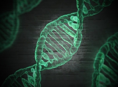
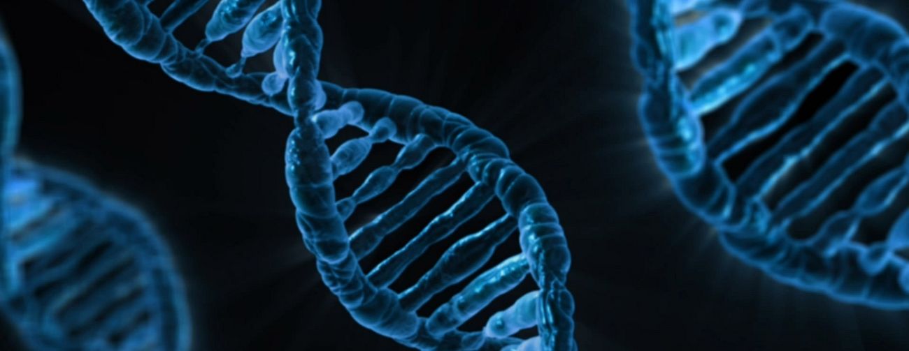
The double helix is the intertwined structure of DNA, enabling genetic information storage, replication, and individual identification, which is crucial in forensic science.
The term “Double Helix” refers to the iconic and remarkable structure of DNA (Deoxyribonucleic AcidDNA, or Deoxyribonucleic Acid, is the genetic material found in cells, composed of a double helix structure. It serves as the genetic blueprint for all living organisms. More), which is central to the fields of genetics, molecular biology, and forensic science. DNA’s double helical structure is a critical aspect of its function, and understanding it is essential for comprehending how genetic information is stored, replicated, and transmitted.
Unraveling the Double Helix
The Birth of a Revolutionary Idea
The term “double helix” is synonymous with the groundbreaking discovery of the DNA’s structure, a remarkable achievement attributed to the brilliant minds of James Watson and Francis Crick. Their pivotal paper, published in the journal Nature in 1953, unveiled the now-famous double helical model of DNA, forever changing the landscape of molecular biology.
Before this revelation, the structure of DNA was a subject of great intrigue. Linus Pauling, a distinguished chemist, and his collaborator Robert Corey had erroneously suggested a triple-stranded conformation for DNA. However, the work of Rosalind Franklin and her student Raymond Gosling, who captured the crucial X-ray diffraction image known as “Photo 51,” laid the foundation for Watson and Crick’s groundbreaking model. This iconic image provided insights into DNA’s helical structure and its spacing’s regularity.
Understanding the Structure
The DNA double helix is a biopolymer composed of nucleic acids held together by nucleotides that base pair, forming the structural basis of the DNA molecule. The most common form of this double helix in nature is the B-DNA, characterized by its right-handed twist, featuring around 10 to 10.5 base pairs per helical turn.
The Major and Minor Grooves
The double helix structure is not just aesthetically pleasing but functionally significant. It exhibits two distinct grooves, the major groove and the minor groove. These grooves play a vital role in molecular interactions.
In B-DNA, the major groove is wider than the minor groove. This variation in groove widths has profound implications. Many proteins that interact with DNA tend to prefer the wider major groove due to its accessibility. The structural nuances of the major and minor grooves are integral to the DNA’s function.
DNA in Various Conformations
DNA is a versatile molecule that can adopt various conformations based on the context and surrounding conditions. While B-DNA is the predominant form in living cells, other conformations like A-DNA and Z-DNA exist.
A-DNA
A-DNA exhibits distinct geometric and dimensional differences compared to B-DNA. It is known to occur in dehydrated samples of DNA, and surprisingly, it has biological functions. A-DNA can be formed when segments of DNA are methylated for regulatory purposes.
Z-DNA
Z-DNA is characterized by the opposite direction of strand winding compared to A-DNA and B-DNA. This unique geometry is observed in regions of DNA where the strands turn about the helical axis differently. Furthermore, protein-DNA complexes can induce the formation of Z-DNA structures.
Other DNA Conformations
Beyond A-DNA, B-DNA, and Z-DNA, various other DNA structures have been created synthetically but are not observed in naturally occurring biological systems. These include C-DNA, E-DNA, L-DNA, P-DNA, and S-DNA.
DNA Bending and Persistence Length
DNA exhibits a certain degree of rigidity, often likened to a worm-like chain. It has three crucial degrees of freedom: bending, twisting, and compression. Bending and twisting play significant roles in various biological processes. The rigidity of DNA segments is typically characterized by the persistence length, which is the length of the polymer segment over which the orientation of the polymer becomes uncorrelated.
The persistence length is essential in understanding the entropic flexibility of DNA under tension. In an aqueous solution, the average persistence length is about 46 to 50 nanometers, equivalent to 140 to 150 base pairs. This measurement indicates that DNA is moderately stiff. However, the persistence length can vary depending on the DNA sequence due to differences in base stacking energies and interactions with the minor and major grooves.
Models for DNA Bending
DNA bending, twisting, and stretching are vital for various cellular processes. At a large scale, DNA behaves according to the worm-like chain model, which describes its bending flexibility. The model is consistent with Hooke’s law for very small forces.
DNA prefers bending in specific directions, often referred to as anisotropic bending. This preference is determined by the stability of stacking each base on top of the next. Bases that cause unstable stacking steps on one side of the DNA helix induce the DNA to bend away from that direction. For instance, A and T residues are typically found inside bends. DNA sequences with exceptional bending preferences can become intrinsically bent. These regions are usually enriched with stretches of A and T residues.
Stretching DNA
Under tension, DNA behaves in an entropically elastic manner. The persistence length and entropic stretching behavior are crucial in understanding how DNA stretches. Longer DNA stretches tend to be entropically elastic, with DNA molecules adopting more compact, relaxed states in solution. This entropic behavior aligns with the Kratky-Porod worm-like chain model and Hooke’s law for small forces.
Phase Transitions Under Stretching
Stretching DNA under tension can lead to phase transitions, resulting in various DNA structures. One proposed structure is the P-form DNA, where the bases splay outward, and the phosphates move to the middle. Additionally, S-form DNA structures have been suggested, characterized by partially melting the base-stack while preserving base-base association. The “Σ-DNA” form, with periodic fracture of the base-pair stack, has also been proposed.
Supercoiling and Topology
DNA molecules in cells are typically found in closed loops or long molecules that effectively form topologically closed domains. DNA can be positively or negatively supercoiled when subjected to torsional strain. Negative supercoiling is prevalent in living cells and aids DNA unwinding for RNA transcription processes.
The Linking Number Paradox
The interplay of linking numbers, twisting, and writhing is crucial in understanding DNA supercoiling and topology. Topoisomerases, a class of enzymes, are responsible for unknotting topologically linked DNA strands, enabling vital processes like DNA replication and recombination to occur.
The double helix, as elucidated by Watson and Crick, stands as one of the most iconic structural discoveries in science. This intricate molecular structure governs the storage and replication of genetic information, playing a pivotal role in the functioning of living organisms. As our understanding of DNA’s complexities deepens, it continues to shape the forefront of biological research and medical advancements, further unraveling the secrets of life.


