

Forensic mitochondrial DNA DNA, or Deoxyribonucleic Acid, is the genetic material found in cells, composed of a double helix structure. It serves as the genetic blueprint for all living organisms. Read Full Definition (mtDNA) interpretation is not simply a matter of reading a DNA sequence and asking whether it matches a known person. Unlike nuclear STR profiles, mitochondrial sequences follow predominantly maternal inheritance, can vary among tissues within a single individual, and are often shared by many people. Their evidential value depends on the sequence region examined, the quality of the results, the comparison criteria, the relevant population data
 Information in analog or digital form that can be transmitted or processed. Read Full Definition and the propositions being considered.
- 1. Why mtDNA can succeed when nuclear STR profiling fails
- 2. Control-region typing versus whole-mitogenome sequencing
- 3. The biological meaning of heteroplasmy
- Worked example 1: calculating a point-heteroplasmy read fraction
- Figure 1: a conceptual heteroplasmy threshold graphic
- 4. Sequence nomenclature: rCRS, RSRS and alignment
- 5. How should an mtDNA sequence comparison be interpreted?
- Worked example 2: heteroplasmy versus a supported exclusion
- 6. Haplogroups: useful phylogeny, not individual identity
- Figure 2: why matching mtDNA cannot uniquely identify someone
- Worked example 3: maternal relatives with the same mitotype
- 7. EMPOP: how forensic mitochondrial database searches work
- 8. From a database count to a frequency and confidence limit
- Worked example 4: one mtDNA match among 500 records
- Figure 3: from mitotype to defensible statistical statement
- 9. What does an mtDNA match mean as forensic evidence?
- 10. NUMTs, mixtures, contamination and analytical pitfalls
- Nuclear mitochondrial sequences (NUMTs)
- Mixed biological contributors versus heteroplasmy
- Contamination and sequence damage
- Sequence homopolymers and platform-dependent errors
- 11. Laboratory validation and interpretation checklist
- 12. How to write a defensible mtDNA report
- 13. Four interpretation errors to avoid
- Frequently asked questions
- Scientific references and official guidance
- Related Simply Forensic resources
mtDNA analysis is particularly valuable for shed hair shafts, old bones, teeth and other degraded or low-nuclear-DNA specimens. A forensic report may identify an mtDNA sequence as consistent with a reference, report a difference whose significance is uncertain, or exclude a source under validated criteria. It should never imply that a maternal-lineage match uniquely identifies a person. Current guidance also requires careful treatment of point and length heteroplasmy, haplotype nomenclature, phylogenetic checks, EMPOP database searches and frequencyFrequency is a fundamental concept in physics and wave theory. It refers to the number of times a specific point on a wave, such as a crest or trough, passes a fixed reference point in Read Full Definition uncertainty.
This specialized guide goes beyond our existing introductions to mtDNA versus nuclear DNA and mtDNA sequencing technologies. It explains interpretation through four reproducible teaching examples and original scientific figures, based on SWGDAM’s 2024 mtDNA guidelines, the ISFG DNA Commission recommendations, official EMPOP documentation and recent forensic validationValidation, often referred to as method validation, is a crucial process in the laboratory when introducing a new machine, technology, or analytical technique. It involves a series of systematic steps and assessments to ensure that Read Full Definition research.
KEY LEARNING POINTS
- mtDNA follows predominantly maternal transmission; a matching mitotype may be shared by maternal relatives and unrelated people.
- Point heteroplasmy and length heteroplasmy are different phenomena, and their interpretation depends on validated methods and sample quality.
- A single sequence difference is not an automatic exclusion. SWGDAM 2024 requires laboratories to establish and document comparison criteria.
- An mtDNA haplogroup is a phylogenetic lineage classification, not a unique identity or definitive ethnicity or birthplace.
- EMPOP searches must use appropriate sequence coverage and population data; a database frequency is not the probability that the reference person was the donor.
1. Why mtDNA can succeed when nuclear STR profiling fails
The human mitochondrial genome A genome is the complete set of an organism's genetic material, while genomics is the study of genomes, investigating their evolution, structure, and function. Read Full Definition is a circular DNA moleculeA molecule is a fundamental unit of matter composed of two or more atoms that are chemically bonded together. It is the smallest possible amount of a particular substance that retains all of the unique Read Full Definition approximately 16,569 base pairs long. It encodes genes
 Genes are DNA segments (or RNA in some viruses) that dictate cellular processes, traits, and hereditary information, promoting genetic diversity and evolution in living organisms. Read Full Definition involved in mitochondrial function and contains a non-coding control region, historically the main target of forensic sequencing. Many cells contain numerous mitochondria and multiple mtDNA molecules per mitochondrion. Consequently, mtDNA can remain available when the two nuclear copies of a locus
 A locus is the precise position of a gene on a chromosome. Different alleles of the same gene occupy the same locus. Read Full Definition are lost, highly fragmented or present in insufficient amounts for a conventional autosomal STR profile.
This copy-number advantage has practical limitations. It varies by tissue, cell type, age and preservation, and higher abundance does not guarantee a clean or complete sequence. A hair shaft may provide useful mtDNA without a satisfactory nuclear profile; a highly damaged bone may require small overlapping targets, special extraction procedures and repeat examinations. Conversely, modern optimized nuclear DNA or SNP methods may sometimes be more informative than mtDNA, especially when individual identification is the principal question.
The laboratory’s choice depends on available specimen, recovery expectations, destructive-sampling considerations and the intended comparison. Our guide to DNA extraction from bones, teeth and degraded remains explains the pre-analytical challenges. An mtDNA analysis from a hair, bone or tooth does not by itself establish when the item was deposited or whether the deposition occurred during the alleged Alleged is a term used in legal contexts to describe an accusation or claim that has been made against an individual but has not yet been proven or confirmed. Read Full Definition event.
2. Control-region typing versus whole-mitogenome sequencing
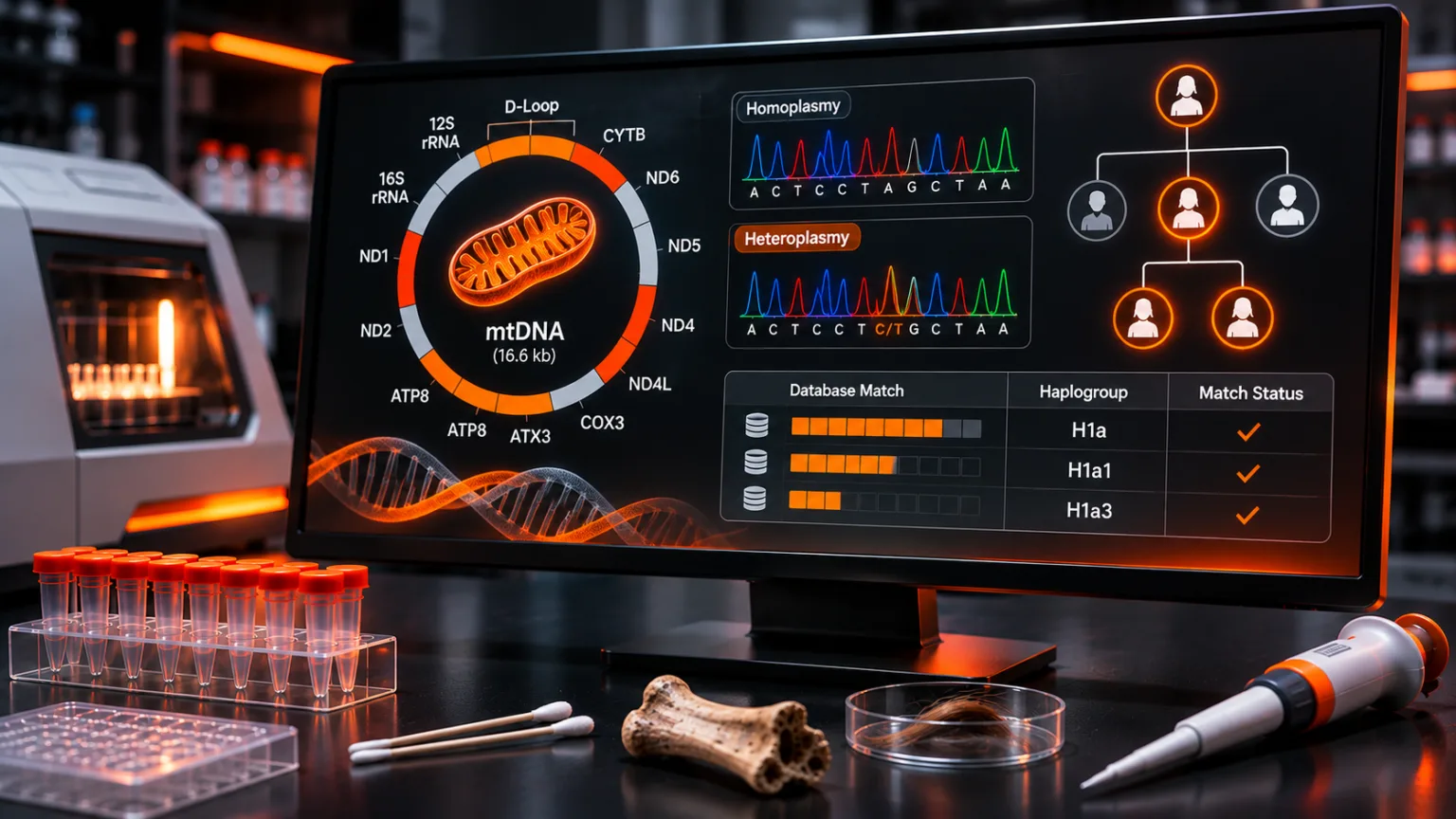
Traditional forensic mtDNA sequencing primarily evaluates segments of the control region, including hypervariable segments HVS-I and HVS-II. These regions contain frequent polymorphisms and historically supported Sanger-based analysis of difficult specimens. Whole-mitogenome sequencing examines the approximately 16.6 kb circular molecule and can resolve some common control-region haplotypes by adding informative coding-region substitutions.
The increased resolution is not universal. Full mtGenome results require adequate coverage across the genome and a suitably comparable population database. The reference database for control-region haplotypes is larger than the complete-mitogenome subset, which can affect how confidently a rare sequence frequency is estimated. Partial specimens may cover different ranges, and counting records that do not actually cover the compared positions is inappropriate. The SWGDAM 2024 guidance addresses how laboratories should choose and document search ranges when comparing whole-mitogenome data with reference databases.
| Approach | Principal data | Strength | Interpretation limitation |
|---|---|---|---|
| Control-region Sanger | Overlapping sequence reads of validated control-region segments | Established method; useful for short targeted regions | Common haplotypes may lack discrimination; low-level variants can be difficult to resolve |
| Control-region MPS/NGS | Numerous sequence reads across selected targets | Can assess minor variant fractions with method-dependent sensitivityIn the context of laboratory equipment or analytical techniques, the term "sensitive" describes the capability of a machine or method to detect even very small amounts or concentrations of a substance. Sensitivity is a quantitative Read Full Definition | Read biases, polymerase errors and alignment of length variants require validation |
| Whole-mitogenome MPS | Control and coding-region positions across approximately 16.6 kb | May distinguish sequences that are identical in the control region | Coverage gaps, NUMTs, degradation and smaller comparable population datasets matter |
Sanger and MPS results should not be compared without aligning their reliably covered positions and recognizing differing detection thresholds. The EDNAP collaborative heteroplasmy study, published online July 23, 2026, found broadly comparable point-heteroplasmy detection but more variability in length-heteroplasmy interpretation, especially in certain homopolymeric regions and sequencing platforms. It provides a strong reminder that increased read depth does not eliminate all methodological differences.
3. The biological meaning of heteroplasmy
Homoplasmy means that the mtDNA molecules evaluated at a specified site are represented by one nucleotide state within the assay’s detection limits. Heteroplasmy means more than one mtDNA sequence variant is present within a person or biological sample. In forensic work, two important forms are distinguished:
- Point heteroplasmy (PHP): two or more nucleotide states occur at the same sequence position—for example, a combination of C and T at a position conventionally written as the IUPAC ambiguity code Y.
- Length heteroplasmy (LHP): mtDNA molecules differ in the number of repeated bases or units, often in homopolymeric C-stretches, producing alternative sequence lengths and potentially complex chromatograms.
Different mtDNA sequence variants can coexist at different proportions within tissues. Their measured ratios may change with sampling, biological drift, cell lineage, age, tissue-specific selection and the mitochondrial genetic bottleneck across generations. A minor variant detectable in hair may be absent or below threshold in blood from the same person. Evidence Evidence is any form of proof, such as objects, materials, or scientific findings, presented to establish or disprove a fact in a legal proceeding. It is used to reconstruct events and link or exclude individuals Read Full Definition of such variation is discussed by Naue and colleagues and more recently by Fisher and colleagues (2026).
Crucially, heteroplasmy must not be assumed every time a minor peak or low-count variant appears. Sequencing errors, PCR artefacts, damage, cross-contaminationCross-contamination - The unwanted transfer of material between two or more sources of physical evidence. For example, improperly collecting biological evidence such as blood could lead to one sample mixing with another sample and contaminating Read Full Definition, NUMT amplification and mixed biological contributions can mimic it. SWGDAM 2024 requires the laboratory to define heteroplasmy within its operational range, set validation-supported base-calling criteria, and validate mtDNA mixture interpretation separately when that is offered.
What thresholds should laboratories use?
There is no universal forensic mtDNA minor-variant threshold appropriate for every sequencing platform, locus and workflow. Studies may use nominal percentages such as 5% or 10% to describe their assays, but a percentage cannot be borrowed from another laboratory without validation. A reportable MPS minor allele should be supported by adequate depth, sufficient independent template representation, expected error performance, base quality and strand or directional checks where applicable. Sanger interpretation relies on appropriate electropherogram quality and confirmed secondary-peak detection criteria rather than simply applying an MPS read-percentage cut-off.
Raw read fractions must also be interpreted cautiously. Two hundred sequencing reads do not necessarily represent two hundred independent starting mtDNA molecules: PCR duplicates, target enrichment and stochastic sampling can affect the estimate. A reported 15% read fraction is a measured proportion of retained reads under the pipeline, not a precise"Precise" refers to the degree of closeness or consistency between multiple measurements or values taken under the same conditions. It indicates how well these measurements agree with each other, regardless of whether they are accurate Read Full Definition measurement of the biological fraction of mitochondria that carry the mutation.
Worked example 1: calculating a point-heteroplasmy read fraction
Consider a purely hypothetical MPS result at position X. After validated read filtering, there are 200 accepted reads: 170 support C and 30 support T. Assume the laboratory’s independently validated minimum variant fraction for reporting at that position is 10%, and all quality, contaminationContamination - The unwanted transfer of material from another source to a piece of physical evidence. The inadvertent touching of a weapon, thereby adding fingerprints to it is an example of evidence contamination. Read Full Definition and biasThe difference between the expectation of the test results and an accepted reference value. Read Full Definition checks pass.
Illustrative read calculation
C fraction = 170 / 200 = 85%
T fraction = 30 / 200 = 15%
15% is above the assumed 10% reporting cut-off. If independently confirmed, the site can be represented as C/T point heteroplasmy (IUPAC Y) under this hypothetical SOP.
That conclusion is contingent on all other validated checks; a single number alone cannot establish true heteroplasmy. If the applicable validated cutoff had instead been 20%, the same minor fraction would fall below that particular reporting threshold. Its omission from a final sequence call would not demonstrate biological absence of the variant.
Figure 1: a conceptual heteroplasmy threshold graphic
POINT HETEROPLASMY · ORIGINAL TEACHING DIAGRAM
200 hypothetical reads
Minor T allele
15%
Above the example 10% threshold, if validation and quality controls are satisfied
A reporting cut-off is a decision rule, not proof of biological absence below that line. MPS read depth, errors, duplicates and contamination remain relevant.
4. Sequence nomenclature: rCRS, RSRS and alignment
Forensic mtDNA haplotypes are commonly described as differences from the revised Cambridge Reference Sequence (rCRS). A notation such as “263G” identifies an observed G at rCRS position 263 when the reference base differs. A homoplasmic position identical to the rCRS is typically omitted from a difference list. Heteroplasmic bases can be shown by IUPAC codes where supported, and insertions or deletions have specific nomenclature conventions. An rCRS-based difference list is not the same as the literal DNA sequence.
The Reconstructed Sapiens Reference Sequence (RSRS) represents a proposed ancestral mtDNA reference for phylogenetic description. The same biological sequence will have a different list of reported differences when compared with RSRS rather than rCRS. Analysts must harmonize the reference framework before comparing notation: contrasting two difference lists built against different references can create apparent mismatches that do not exist in the underlying nucleotides.
Alignment is especially difficult in repeated bases. The 2024 SWGDAM document favors a phylogenetic alignment approach verified using EMPOP and specifies rules for indels and common homopolymer tracts. Such rules help prevent identical underlying strings from being incorrectly treated as distinct haplotypes because a laboratory placed an insertion or deletion at a different repeated position.
Forensic software may also evaluate haplogroup-defining mutations to flag implausible or missing sites and possible sequence transcription mistakes. The 2014 ISFG guidance provides foundational nomenclature and phylogenetic quality-control principles; the SWGDAM 2024 revision extends their practical treatment for both Sanger and MPS data.
5. How should an mtDNA sequence comparison be interpreted?
A comparison should begin with the overlapping, reliable sequence range and the laboratory’s validated quality requirements. An exact match in the comparable region generally supports a cannot-exclude outcome, although its evidential weight depends on the population frequency of that sequence. If heteroplasmy is present, the analystA designated person who examines and analyzes seized drugs or related materials, or directs such examinations to be done; independently has access to unsealed evidence in order to remove samples from the evidentiary material for Read Full Definition must follow approved rules that account for possible differences between tissues or across maternal transmissions. Length variation at common poly-C tracts may be especially unhelpful for deciding whether two samples differ in source.
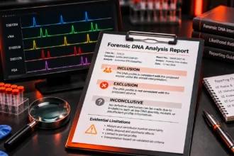
The most important caution is that one base difference is not universally defined as an exclusion. SWGDAM’s 2024 guidelines, section 5.1, require each laboratory to define conditions for inclusion, exclusion and inconclusive outcomes. They advise establishing the number of differences justifying exclusion using published and/or internal research, taking account of sequenced region, detection thresholds and whether the comparison concerns a direct source or more distant maternal relatives. Historically, some laboratories have treated a single unambiguous difference as inconclusive and multiple differences as exclusionary; those historical conventions should not be presented as a universal modern rule.
Equally, the term “cannot exclude” does not prove that the evidence originated from the reference person. Maternal relatives can share a type, and other people without a known genealogical relationship may carry the same sequence. The outcome of a valid mtDNA comparison is distinct from the question of when or how material reached the surface.
Worked example 2: heteroplasmy versus a supported exclusion
Assume that evidence and reference samples were sequenced across the same high-quality set of positions, using a method that has valid contamination and heteroplasmy controls. Consider three synthetic situations:
| Scenario | Reference | Evidence | Interpretation under stated assumptions |
|---|---|---|---|
| A: exact agreement | 16024–16365 region, selected sites all concordant | Same nucleotide states at every comparable site | Cannot exclude the reference or another matching maternal-lineage source |
| B: point heteroplasmy | Position X: homoplasmic C | Position X: C/T, high-quality heteroplasmy; all other positions concordant | Potentially compatible under a validated heteroplasmy rule; not automatically exclusion |
| C: multiple stable differences | Positions X, Y, Z: A, C, G | Positions X, Y, Z: G, T, A, with no heteroplasmy detected and all calls reproducible | May justify exclusion as a direct source if the laboratory’s validated rule and relevant biological context support it |
Scenario B is not equivalent to a novel third contributor merely because two bases are present in the sample. Scenario C does not mean every distant maternal relationship is impossible: its conclusions depend on the specified relationship, sequence range, mutational processes and laboratory rules. A single stable homoplasmic difference between a hair and a blood reference may warrant additional sampling or an inconclusive comparison, rather than immediate categorical exclusion.
Length heteroplasmy introduces its own issues. For example, alternative numbers of C bases following a common control-region anchor can be observed in material from the same person. The 2024 SWGDAM guidance does not recommend using certain common length variants, including those following positions 16193, 309 and 573, for comparisons or searches. Specific exceptions and quality procedures should be documented.
6. Haplogroups: useful phylogeny, not individual identity
An mtDNA haplogroup is a branch of the maternal phylogenetic tree defined by a pattern of mutations inherited over many generations. Broad clades such as L, M, N and H contain numerous subclades and many people. A haplogroup classification may support quality controlQuality control (QC) refers to a series of activities and measures conducted on individual laboratory tests or analyses to verify and ensure the accuracy and reliability of the results. QC is a reactive approach that Read Full Definition and historical population research, but it does not identify one person, uniquely indicate a nationality, or establish a person’s ethnic identity or birthplace.
Tools such as HaploGrep 3 assign mitotypes to phylogenetic lineages using a selected tree and scoring procedure. The 2023 HaploGrep 3 publication describes its classification and quality-control functions. Reproducibility requires documenting the actual software and phylogeny version, for example a specified PhyloTree build or forensic update. The EMPOP service has also evolved: its current documentation describes mitoLEAF as part of its haplogrouping workflow from 2025. These references are related phylogenetic resources, not interchangeable software settings.
A surprising haplogroup assignment can provide a valuable signal to investigate variant calling, dropout, alignment, sample mix-ups or contamination. It is not by itself proof that the sample must have been collected from a particular ethnic group. In populations with extensive migration and historical intermixing, a maternal clade’s geographic distribution can overlap many present-day countries and communities.
Figure 2: why matching mtDNA cannot uniquely identify someone
MATERNAL INHERITANCE · ORIGINAL SIMPLY FORENSIC DIAGRAM
Grandmother
Maternal mitotype H
Mother
H, allowing mutation
Son
Receives maternal mtDNA
Daughter
Receives and can transmit mtDNA
Both sons and daughters normally inherit mtDNA from their mother. Daughters can transmit their mitochondrial genome to the next generation; sons generally do not. Sequence differences and variant proportions can arise through mutation and heteroplasmy.
Worked example 3: maternal relatives with the same mitotype
A mother, her daughter and her son each provide high-quality reference samples. Their interpreted sequences at all 500 reliably compared positions are identical. A crime-scene hair shaft matches these positions. The mitochondrial evidence is therefore consistent with any of these three people, subject to the applicable laboratory’s comparison rules, as well as other people sharing the same haplotype. The three individuals are distinct people even though their mtDNA profiles match.
Now suppose the daughter has a verified minor T variant at one position, representing 8% of high-quality reads, while the mother and son are reported as C at the same site. That difference may reflect heteroplasmy distribution, tissue sampling or detection limits. It does not automatically establish that the daughter is or is not the donor. An exact statistical probability of their relationship or relative source contributions cannot be calculated from these statements alone.
This example also demonstrates why a rare mtDNA haplotype does not exclude maternal relatives as alternative sources. Population rarity describes the type in a sample of people; it does not transform a shared maternal sequence into an individual-identification marker.
7. EMPOP: how forensic mitochondrial database searches work
EMPOP, the EDNAP mitochondrial DNA population database, provides quality-controlled reference sequences and database searches for forensic comparisons. It is not a comprehensive census of all maternal lineages. Dataset composition, geographic coverage, sequence ranges and the release being consulted matter. At the time of the current official query documentation, EMPOP Release 14 distinguishes substantially different numbers of control-region and complete-mitogenome records. A user cannot assume every record provides the same sequence coverage.
EMPOP uses an alignment-free string approach (SAM2) to compare equivalent sequence strings despite potential differences in their position-based indel representation, and supports phylogenetic alignment and haplogroup quality control. The original String-based Sequence Search Algorithm was described by Röck and colleagues (2011); current EMPOP documentation describes the later SAM2 workflow. Do not treat a simple text search for a mutation list as equivalent to a validated alignment-aware or alignment-free forensic database query.
Step-by-step EMPOP workflow
- Establish the consensus: review sequence quality, interpretable regions, heteroplasmy and any ambiguous sites against the validated SOP.
- Normalize nomenclature: record rCRS differences consistently and check the underlying sequence/phylogenetic alignment.
- Specify coverage: distinguish HVS-I, HVS-I/HVS-II, complete control region, partial sequences and complete mtGenome records.
- Choose a relevant reference set: document the population or metapopulation, its size and why it is appropriate for the case.
- Inspect the search: record exact matches, available comparable records, sequence ranges, ambiguous states and selected search parameters.
- Compute evidential weight: distinguish the observed proportion from the chosen conservative confidence bound or other justified method.
- Retain reproducibility information: EMPOP release, access date, query specifications, output and any alternative search decisions.
A full-mitogenome case sequence searched against a control-region-only reference may generate many more apparent matches than the full sequence would. Conversely, excluding records with less sequence coverage reduces the reference sampleReference sample - material from a verifiable/documented source which, when compared with evidence of an unknown source, shows an association or linkage between an offender, crime scene, and/or victim. Read Full Definition size, increasing sampling uncertainty. SWGDAM 2024 explicitly addresses reduced-range searches: the search strategy must not count shorter-range matches that would be excluded by the longer reliable range being evaluated.
Population inference needs care. A country label is not an exhaustive descriptor of maternal ancestry, and a geographically small or historically founded population may have different frequencies from a large continental reference set. Analyses should not select the lowest available frequency simply because it gives a larger statistical weight.
8. From a database count to a frequency and confidence limit
Let x equal the observed number of matching haplotypes among n comparable database records. The sample frequency is x/n. A laboratory may also calculate an augmented-count estimate such as (x+1)/(n+1), or an upper confidence limit using a binomial approach. These quantities answer different questions. The SWGDAM 2024 guidelines describe counting as the basis of frequency estimation and recommend an upper bound probability estimate incorporating a confidence interval, generally 95% or greater.
| Statistic | Definition | Interpretation |
|---|---|---|
| Observed frequency | x/n | Observed fraction of comparable records, not exact population truth |
| Augmented count | (x+1)/(n+1) | Pseudocount-based point estimate, not a confidence bound |
| One-sided 95% Clopper–Pearson upper limit | Upper p satisfying P(X ≤ x | Binomial(n,p)) = 0.05 | Upper confidence bound under the stated binomial model |
| Two-sided exact 95% interval upper endpoint | Upper p satisfying P(X ≤ x | Binomial(n,p)) = 0.025 | Different endpoint; uses 2.5% upper-tail allocation |
These calculations treat reference entries as if they were independent observations from a specified population. Real-world datasets can be affected by convenience sampling, maternal relatedness, underrepresentation and database composition. Confidence limits quantify the sampling uncertainty under their statistical assumptions; they do not correct all biological or sampling biases.
Worked example 4: one mtDNA match among 500 records
Assume a synthetic control-region haplotype was observed once in a dataset containing n = 500 eligible, comparable mitotypes. The database count is x = 1. We calculate:
Observed frequency: 1/500 = 0.002 = 0.20%
Augmented estimate: 2/501 = 0.003992 = 0.3992%
One-sided exact 95% upper limit: approximately 0.009452 = 0.9452%
Two-sided exact 95% upper endpoint: approximately 0.011092 = 1.1092%
The one-sided and two-sided limits are not identical; they use different tail conventions. In this example, the one-sided upper limit corresponds to the binomial probability of observing one or fewer matches equal to 0.05, given 500 draws. These percentages are hypothetical mathematical results rather than EMPOP search output, and a casework report must state which procedure the laboratory adopted.
Now suppose no matching sequence was observed among the same 500 records (x = 0). The observed frequency is zero, but the one-sided exact 95% upper confidence limit is 1 − 0.05^(1/500) ≈ 0.005974 = 0.5974%. The upper endpoint of a two-sided 95% interval is 1 − 0.025^(1/500) ≈ 0.007351 = 0.7351%. Neither result establishes that the haplotype is unique in the population. Applying a “rule of three” gives an approximate one-sided zero-count upper limit near 3/500 = 0.6%, but it should not replace the exact bound when the latter is available.
Figure 3: from mitotype to defensible statistical statement
EMPOP SEARCH AND REPORTING · ORIGINAL SIMPLY FORENSIC WORKFLOW
1 · Sequence
Validated calls and coverage
2 · Normalize
rCRS and phylogenetic QC
3 · Search
EMPOP cohort, loci, release
4 · Estimate
Count and approved upper limit
5 · Report
Maternal sharing and caveats
No database matches ≠ zero population frequency ≠ unique source identification. Document actual sample size and statistical convention.
9. What does an mtDNA match mean as forensic evidence?
A validated mtDNA sequence comparison may yield a cannot-exclude result, and a population search may quantify how often that sequence occurred within the selected reference records. But P(sequence | a random alternative source) is not the same as P(a particular person is the source | sequence). The latter requires additional prior information and other evidence. The statistical treatment of maternal relatives also differs from that of independent alternative donors.
Under deliberately restricted assumptions—one high-quality haplotype, perfect detection, a specified random unrelated alternative, and an accepted population-frequency estimate—an illustrative source-level likelihood ratio for a matching type could be proportional to the reciprocal of that population frequency. The real forensic question is whether those assumptions are appropriate, whether mutation and heteroplasmy are material, and whether a maternal-lineage relative is a plausible alternative. A reciprocal frequency must not be presented as an automatic, universal mtDNA LR.
This is analogous to the cautions for Y-STR paternal-lineage interpretation, but mtDNA has its own biology, heteroplasmy and technical artefacts. For a fuller explanation of likelihood ratios and their interpretation, see our DNA mixture statistics guide and DNA reporting guide.
10. NUMTs, mixtures, contamination and analytical pitfalls
Nuclear mitochondrial sequences (NUMTs)
Human chromosomes contain fragments of mitochondrial-origin DNA, known as NUMTs. Depending on assay design and specimen characteristics, some can co-amplify or align to the mitochondrial reference and generate misleading variants or apparent heteroplasmy. NUMT interference is not guaranteed in every assay, but it is an important validation consideration, particularly with short fragments and highly sensitive sequencing. Bintz, Dixon and Wilson (2014) investigated detection of mtDNA and nuclear-inserted sequences under forensic amplification strategies.
Mixed biological contributors versus heteroplasmy
Two bases at one position do not by themselves distinguish heteroplasmy from a mixture of biological contributors. A mixed DNA source may show systematic sets of alternative alleles Alleles are different forms of a gene resulting from mutations or variations in the DNA sequence or gene expression. They can be dominant (expressed with one copy) or recessive (expressed only with two copies). Read Full Definition at multiple positions or differing read patterns. However, multiple heteroplasmic sites are also biologically possible, especially in complex tissues. The analyst needs the entire sequence profile, validated thresholds, replicate data, controls, specimen context and where suitable, orthogonal confirmation. If a mixture cannot be resolved, a single-source frequency calculation is not justified.
Contamination and sequence damage
Contamination may occur during collection, handling, extraction, PCR setup, library preparation or sequencing. The high sensitivity of mtDNA typing means low-level extraneous molecules can be especially problematic. Negative controls, elimination comparisons, physically or procedurally separated workflows and validated repeat testing are central quality measures. Chemical damage in degraded remains may also produce characteristic apparent substitutions requiring technical evaluation. If a control contains contamination, the case cannot simply be called valid because the evidence contains a stronger signal; the laboratory must apply its established contamination-assessment policy.
Sequence homopolymers and platform-dependent errors
Repeating C-stretches can generate difficult length patterns. Sanger traces may become mixed after a variable-length stretch; MPS assays can generate PCR-slippage and platform-specific indel errors. The 2026 EDNAP collaborative study is particularly relevant here: it found that length heteroplasmy remained less reproducible between methods than point heteroplasmy, despite standardization efforts. Accordingly, a whole-mitogenome sequence with very high average depth can still contain difficult local positions.
11. Laboratory validation and interpretation checklist
Valid forensic mtDNA interpretation requires more than a successful sequencing run. Under the FBI Quality Assurance Standards effective July 1, 2025, laboratories within scope must validate their methods and define interpretation procedures. The 2024 SWGDAM guidelines distinguish expected control performance, minimum sequence quality, base-call assignment, heteroplasmy definition, mixture validation, nomenclature and casework frequency calculations. These guidelines inform best practice but are not automatically legally binding in every country.
| Validation / QA area | What the laboratory should examine | Interpretive risk addressed |
|---|---|---|
| Sample types and sensitivity | Bone, teeth, hair, blood, aged/degraded samples and dilution series | Unsupported extrapolation beyond validated samples |
| Variant detection | Point-heteroplasmy fraction, strand bias, base quality, depth and replicate behaviour | False minor variants or missed true variants |
| Repeat/homopolymer regions | Length heteroplasmy, slippage, indel alignment and platform artefacts | False differences or incorrect frequency searches |
| NUMTs and specificityIn the context of laboratory equipment or analytical techniques, the term "specific" describes the capability of a machine or method to accurately and selectively detect particular molecules or substances while minimizing interference from others. Specificity Read Full Definition | Primer specificity, mapping ambiguity and off-target nuclear signal | Misassigned mtDNA variants |
| Contamination and mixtures | Reagent blanks, negatives, mixture designs, elimination sources and decision criteria | Misinterpreting mixed or extraneous DNA |
| Computational processing | Software versions, read filters, reference assembly, haplogroup tree and reproducibility | Hidden pipeline-dependent profile changes |
| Reporting and statistics | Comparison categories, database representation, Clopper–Pearson settings and proposition scope | Overstatement of inclusion or rarity |
Records should preserve the relevant raw data, sequences, QC metrics, interpretations, versioned pipelines, population search details and technical review. New versions of analysis software or phylogenetic trees may require appropriate change control. The question is whether the specific method as used reliably supports the issued conclusion.
12. How to write a defensible mtDNA report
Reports should clearly identify the specimen and reference, regions sequenced, level of sequence quality, comparison outcome, any heteroplasmic positions or excluded hotspots, reference population and frequency method. A complete report or accessible case record should document analysis software, population search settings and material limitations where required by applicable policies.
Illustrative wording: compatible mtDNA sequences
“The mitochondrial DNA sequence obtained from questioned specimen Q is consistent with the reference mtDNA sequence of individual R over the interpretable positions compared. Individual R cannot be excluded as a possible source of the mtDNA. Because mitochondrial DNA is inherited along maternal lineages, maternally related individuals and other persons sharing the haplotype also cannot be distinguished by this result. The observed haplotype frequency and the laboratory’s approved confidence-bound estimate are reported with the relevant population dataset, sequence range and search information.”
For incompatible results, explain the number and quality of differences and apply the laboratory’s validated exclusion rules. For unresolved or partial results, identify exactly what prevented a supported comparison. Avoid a blanket claim that a database-zero haplotype can only have come from one individual. Also avoid reporting a mitochondrial haplogroup as an individual identity, country of origin, ethnicity or a reliable description of personal ancestry.
Forensic interpretation is usually at the source or lineage level. Whether the material was transferred by direct contact, secondary transfer, old handling or another activity involves separate evidence and propositions. The DNA report should not silently convert a lineage-level observation into a statement that a person participated in an alleged activity.
13. Four interpretation errors to avoid
- Excluding a person based on one difference without considering heteroplasmy: apply validated comparison criteria; differences among tissue samples and among maternal relatives may occur.
- Calling a low-level MPS variant genuine based only on a read percentage: check errors, DNA damage, mapping, strand support, sample quality and contamination.
- Using a whole-mitogenome haplotype frequency from a control-region-only search: statistics must reflect comparable sequence coverage, counts and population selection.
- Calling a matching mtDNA haplotype an individual identification: maternal relatives and unrelated persons may share the type; even a rare haplotype cannot uniquely identify one person.
Frequently asked questions
What is heteroplasmy in forensic mtDNA analysis?
It is the presence of more than one mitochondrial sequence variant within an individual or sample. Point heteroplasmy concerns base differences; length heteroplasmy concerns variable sequence length. Both require validation-supported interpretation.
Does one mtDNA nucleotide difference exclude someone?
Not automatically. SWGDAM 2024 requires laboratories to define exclusion criteria using evidence, the sequence range, detection limits and relevant relationship. Heteroplasmy and mutation can complicate comparisons.
What is the difference between rCRS and RSRS?
They are different mitochondrial reference sequences. The same biological mtDNA sequence may have different variant lists when described relative to rCRS or RSRS; harmonize reference and alignment before comparison.
Can mtDNA haplogroups identify nationality or ethnicity?
No. Haplogroups describe broad maternal phylogenetic branches, often distributed across many populations. They are not individual identifiers and do not determine nationality or ethnicity.
How does EMPOP search mitochondrial DNA?
EMPOP uses string-based sequence comparison to locate matching mitotypes despite possible differences in alignment notation. Searches must use appropriate sequence ranges, population records and quality controls.
What if an mtDNA haplotype is absent from a database?
The observed count is zero in the chosen sample, not proof of zero population frequency. An appropriate confidence upper limit can express sampling uncertainty under a stated statistical model.
Why is a 95% one-sided confidence limit different from a two-sided one?
They allocate error probabilities differently. With no matches in 500 idealized records, the one-sided exact 95% upper limit is approximately 0.5974%, whereas the upper endpoint of a two-sided exact 95% interval is about 0.7351%.
Can a mother and child have different mtDNA sequences?
Yes. Mitochondrial variants can shift in frequency through maternal transmission and across tissues. Close maternal relatives usually share broadly similar mitotypes, but differences are possible.
Why can NUMTs cause problems in mitochondrial sequencing?
NUMTs are nuclear DNA regions of mitochondrial origin. Under some assay conditions they can amplify or map as mitochondrial reads, mimicking true variants or heteroplasmy. Laboratories must evaluate specificity and computational filtering.
Can an mtDNA match prove that one individual deposited the evidence?
No. Matching mtDNA is consistent with possible maternal-lineage sources and other people sharing the mitotype. The match does not establish unique identity, time, transfer mechanism or alleged activity.
Scientific references and official guidance
View 17 scientific references and authoritative resources
- Scientific Working Group on DNA Analysis Methods. Interpretation Guidelines for Mitochondrial DNA Analysis by Forensic DNA Testing Laboratories. Approved and effective September 9, 2024.
- SWGDAM. Supplemental Information for the SWGDAM Interpretation Guidelines for Mitochondrial DNA Analysis by Forensic DNA Testing Laboratories. 2024.
- Parson W, Gusmão L, Hares DR, et al. DNA Commission of the ISFG: revised and extended guidelines for mitochondrial DNA typing. Forensic Science
 Discover the fascinating field of Forensic Science, the application of scientific principles to legal matters. This post delves into its many disciplines, from DNA analysis to crime scene investigation, its importance in the justice system, Read Full Definition International: Genetics. 2014;13:134–142.
- Ewers L, Huber G, Anslinger K, et al. The EDNAP collaborative study on mtDNA heteroplasmy detection using Sanger and Massively Parallel Sequencing technologies. Published online July 23, 2026; Forensic Science International: Genetics, volume 86 (January 2027), article 103593.
- EMPOP. EDNAP mitochondrial DNA population database. Official online resource and current documentation; record database release and date when reporting results.
- Röck A, Irwin J, Dür A, Parsons T, Parson W. SAM: String-based sequence search algorithm for mitochondrial DNA database queries. Forensic Science International: Genetics. 2011;5:126–132.
- Schönherr S, Weissensteiner H, Kronenberg F, Forer L. Haplogrep 3—an interactive haplogroup classification and analysis platform. Nucleic Acids Research. 2023;51:W263–W268.
- HaploGrep 3. Available phylogenetic trees and version information. Official documentation; accessed for this guide in November 2026.
- Naue J, Hörer S, Sänger T, et al. Evidence for frequent and tissue-specific sequence heteroplasmy in human mitochondrial DNA. Mitochondrion. 2015;20:82–94.
- Fisher N, et al. Mitochondrial genetic variation across tissues of the human body. Nucleic Acids Research. Published online May 25, 2026.
- Bintz BJ, Dixon GB, Wilson MR. Simultaneous detection of human mitochondrial DNA and nuclear-inserted mitochondrial-origin sequences using forensic amplification strategies and pyrosequencing technology. Journal of Forensic Sciences. 2014;59:1064–1073.
- Sturk-Andreaggi K, Peck MA, Boysen C, et al. AQME: A forensic mitochondrial DNA analysis tool for next-generation sequencing data. Forensic Science International: Genetics. 2017;31:189–197.
- FBI. Quality Assurance Standards for Forensic DNA Testing Laboratories. Effective July 1, 2025.
- Clopper CJ, Pearson ES. The use of confidence or fiducial limits illustrated in the case of the binomial. Biometrika. 1934;26:404–413.
- Alqaisi MHM, Ekka MM, Patel BC. Forensic evaluation of mitochondrial DNA heteroplasmy in Gujarat population, India. Annals of Human Biology. 2022;49:332–341.
- EMPOP. Using EMPOP for mitotype searches. Official search and haplogrouping documentation, including recent phylogeny developments.
- National Institute of Standards and Technology. Effective strategies for forensic analysis in the mitochondrial DNA coding region. Official research publication.

Related Simply Forensic resources
- mtDNA vs Nuclear DNA: Why Size and Inheritance Matter
- Short-Read vs Long-Read mtDNA Sequencing
- DNA Extraction from Bone, Teeth and Degraded Remains
- Y-STR Interpretation: Paternal-Lineage Limitations
- Forensic DNA Reporting: Inclusion, Exclusion and Inconclusive Results
- DNA Mixture Statistics: CPI, RMP and Likelihood Ratios